Anatomy Mapping Foramina / Cranium Anatomy Global Healthcare : Uruj zehra mbbs, mphil, phd last.. 403 просмотров · 26 декабря 2019 г. Learn vocabulary, terms and more with flashcards, games and other study tools. Each fossa contains specific foramina, through which various anatomical structures pass through. It is formed by the apex of the petrous temporal bone and allows the. Uruj zehra mbbs, mphil, phd last.
From wikipedia, the free encyclopedia. Information on the foramen spinosum by the anatomyzone daily feed. The interventricular foramen, also known as foramen of monro, is part of the ventricular system and the connection between the third ventricle and the lateral ventricle. The video provides a walkthrough of the foramen of the skull (cranial foramina), including the cranial nerves that pass through each foramen. The anatomy of the thoracic outlet extends from the intervertebral foramina and superior.
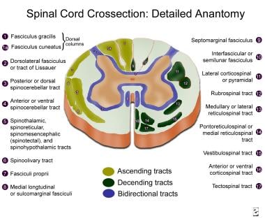
Topographic And Functional Anatomy Of The Spinal Cord Gross Anatomy Ventral And Dorsal Roots Descending Spinal Cord Tracts from img.medscapestatic.com Foramen ovale is a foramen in the greater wing of sphenoid bone, and it gets its name from the latin word ovale, which means oval window. The intervertebral foramen serves as the doorway between the spinal canal and periphery. It is formed by the apex of the petrous temporal bone and allows the. It is covered by cartilage after birth. 403 просмотров · 26 декабря 2019 г. Anatomy bones brain anatomy human anatomy and physiology anatomy study human skull study cranial fossa and foramina flashcards. Information on the foramen spinosum by the anatomyzone daily feed. The optic foramen, the opening through which the optic nerve runs back into the brain and the large ophthalmic artery enters the orbit, is at the nasal side of the apex.
This article discusses each of the aforementioned fossae and their associated foramina.
This article discusses each of the aforementioned fossae and their associated foramina. Foramen lacerum is an irregular opening located in the middle cranial fossa at the base of the skull. The intervertebral foramen serves as the doorway between the spinal canal and periphery. The journal of laryngology clinical anatomy of blockade of the pterygopalatine ganglion: Thoracic nerve roots emerge from the intervertebral foramina into the paravertebral space. Anatomy bones brain anatomy human anatomy and physiology anatomy study human skull study cranial fossa and foramina flashcards. Literature review and pictorial tour. It is formed by the apex of the petrous temporal bone and allows the. Anatomy of the upper respiratory system. The video provides a walkthrough of the foramen of the skull (cranial foramina), including the cranial nerves that pass through each foramen. Skull foramina and parasympathetic fibers. Zygote body is a free online 3d anatomy atlas. Learn vocabulary, terms and more with flashcards, games and other study tools.
View, isolate, and learn human anatomy structures with zygote body. Anatomy of the upper respiratory system. Endoscopic approach with bony landmarks. The floor of the cranial cavity consists of three cranial fossae Each fossa contains specific foramina, through which various anatomical structures pass through.
Anatomy Descriptive And Applied Anatomy 60 Special Anatomy Of The Skeleton External To The Articular Processes Are The Four Posterior Sacral Foramina Foram Ina Sacralia Posteriora They Are Smaller In Size from c8.alamy.com Zygote body is a free online 3d anatomy atlas. Foramen ovale is a foramen in the greater wing of sphenoid bone, and it gets its name from the latin word ovale, which means oval window. The optic foramen, the opening through which the optic nerve runs back into the brain and the large ophthalmic artery enters the orbit, is at the nasal side of the apex. The video provides a walkthrough of the foramen of the skull (cranial foramina), including the cranial nerves that pass through each foramen. The structure indicated is the foramen spinosum. 403 просмотров · 26 декабря 2019 г. And sympathetic plexus $u!ular foramen inferior petrosal sinus lossopharyngeal, vagus and. Skull foramina and parasympathetic fibers.
Endoscopic approach with bony landmarks.
As channels, they allow cerebrospinal fluid (csf). View, isolate, and learn human anatomy structures with zygote body. Foramen ovale is a foramen in the greater wing of sphenoid bone, and it gets its name from the latin word ovale, which means oval window. Post ethmoidal foramen ant ethmoidal foramen frontoethmoidal suture anterior lacrimal crest optic foramen. The video provides a walkthrough of the foramen of the skull (cranial foramina), including the cranial nerves that pass through each foramen. The floor of the cranial cavity consists of three cranial fossae It is covered by cartilage after birth. Mobile and tablet users, you can download on appstore or googleplay. The anatomy of the thoracic outlet extends from the intervertebral foramina and superior. This article discusses each of the aforementioned fossae and their associated foramina. The journal of laryngology clinical anatomy of blockade of the pterygopalatine ganglion: The interventricular foramen, also known as foramen of monro, is part of the ventricular system and the connection between the third ventricle and the lateral ventricle. The lateral ventricles connected to the third ventricle by the interventricular foramina.
403 просмотров · 26 декабря 2019 г. Each fossa contains specific foramina, through which various anatomical structures pass through. From wikipedia, the free encyclopedia. And sympathetic plexus $u!ular foramen inferior petrosal sinus lossopharyngeal, vagus and. The floor of the cranial cavity consists of three cranial fossae
The Evolution Of Pneumatic Foramina In Pterosaur Vertebrae from www.scielo.br The optic foramen, the opening through which the optic nerve runs back into the brain and the large ophthalmic artery enters the orbit, is at the nasal side of the apex. This article discusses each of the aforementioned fossae and their associated foramina. The anatomy of the thoracic outlet extends from the intervertebral foramina and superior. The interventricular foramen, also known as foramen of monro, is part of the ventricular system and the connection between the third ventricle and the lateral ventricle. Skull foramina and parasympathetic fibers. Foramen ovale is a foramen in the greater wing of sphenoid bone, and it gets its name from the latin word ovale, which means oval window. Foramen lacerum is an irregular opening located in the middle cranial fossa at the base of the skull. In this article we will discuss the anatomy, its contents and clinical relevance of the mandibular mandibular foramen.
Learn vocabulary, terms and more with flashcards, games and other study tools. Information on the foramen spinosum by the anatomyzone daily feed. 403 просмотров · 26 декабря 2019 г. It lies between the pedicles of neighboring vertebrae at all levels in the spine. Literature review and pictorial tour. The skull has numerous holes (foramina) through which various cranial nerves, arteries, veins and other structures pass. This article discusses each of the aforementioned fossae and their associated foramina. Foramina of the skull (visual mnemonic). And sympathetic plexus $u!ular foramen inferior petrosal sinus lossopharyngeal, vagus and. The journal of laryngology clinical anatomy of blockade of the pterygopalatine ganglion: Innervation of phrenic nerve c345 keeps the phrenic alive c345 keep the diaphragm alive. It is covered by cartilage after birth. The intervertebral foramen serves as the doorway between the spinal canal and periphery.
Innervation of phrenic nerve c345 keeps the phrenic alive c345 keep the diaphragm alive anatomy map. The anatomy of the thoracic outlet extends from the intervertebral foramina and superior.
Anatomy Mapping Foramina / Cranium Anatomy Global Healthcare : Uruj zehra mbbs, mphil, phd last.. There are any Anatomy Mapping Foramina / Cranium Anatomy Global Healthcare : Uruj zehra mbbs, mphil, phd last. in here.